

Flow cytometry is an analytical technique that measures the physical and chemical properties of individual cells or particles as they pass in a single file through one or more laser beams in a fluid stream. Each cell scatters light and, depending on which fluorescent markers it carries, emits fluorescence that a detector array converts into data on cell size and internal structure, along with specific protein expression. A single flow cytometry run can characterize thousands of cells per second, giving researchers a level of single-cell resolution that bulk assays cannot match.
Every flow cytometry run rests on three measurements collected as each cell crosses the laser.
Light that bends slightly around the cell in the same direction as the laser beam. FSC intensity correlates with cell size, so larger cells produce a stronger forward scatter signal.
Light that deflects at roughly a 90-degree angle off internal cell structures. SSC intensity reflects granularity and internal complexity, which is why granulocytes produce a much higher SSC signal than lymphocytes.
Light emitted by fluorochromes conjugated to antibodies or other fluorescent probes bound to a cell-surface or intracellular antigen. Each fluorochrome emits at a characteristic wavelength, and the instrument's detectors sort that emitted light by color to identify which markers a given cell carries.
Plotting FSC against SSC gives researchers a first-pass view of a sample's cell populations by size and granularity alone, before a single fluorescent marker is added. Layering fluorescence data on top of that scatter profile measures the specific protein markers each individual cell expresses, at the same time and on the same cell.
A flow cytometer runs on three integrated systems working together in real time.
The sample enters the instrument in suspension and passes through a narrow flow cell, where the sheath fluid focuses the cells into a single-file stream via hydrodynamic focusing. This step is what allows the instrument to interrogate one cell at a time rather than a clump.
As each cell crosses one or more laser beams, it scatters light and, if labeled, emits fluorescence. A series of lenses and mirrors, paired with optical filters, directs specific wavelengths of that light toward dedicated detectors. Forward scatter and side scatter each land on their own detector, and every fluorescence channel gets measured separately as well.
Photodetectors convert the collected light into electronic signals, which get digitized and recorded as a data point for that individual cell. Across a full run, this produces a dataset where every row represents one cell and every column represents one measured parameter, ready for gating and analysis in flow cytometry software.
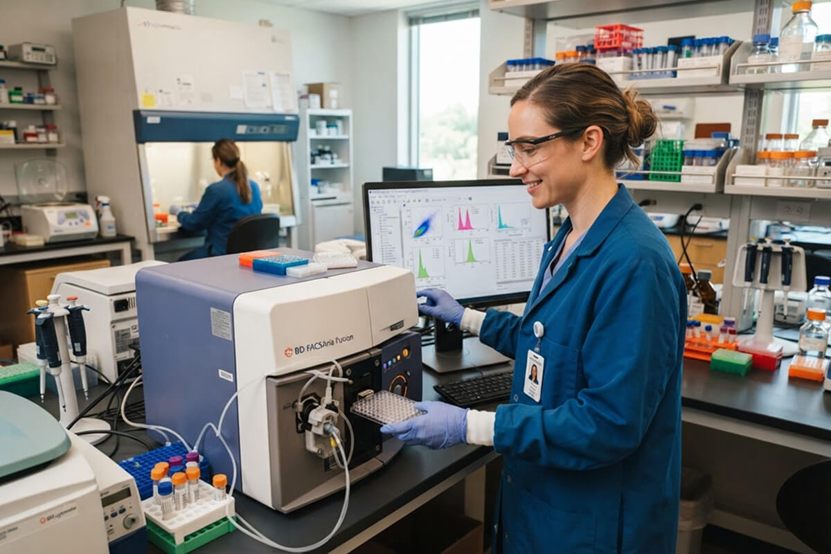
Standard flow cytometry is an analysis-only process. The instrument measures and records data for each cell as it passes through the laser, but the cell then continues to waste.
Fluorescence-activated cell sorting (FACS) adds a physical separation step on top of that same measurement process: after a cell is measured, the instrument applies an electrical charge and deflects it into a specific collection tube based on which markers it expresses. In practice, this means every FACS run is a flow cytometry run, but not every flow cytometry run sorts cells.
Researchers who need to isolate a live, viable population, such as a specific T cell subset for downstream culture, need a cell sorter. Researchers who only need data, such as the proportion of a marker-positive population in a sample, can run standard flow cytometry analysis.
Flow cytometry supports applications across immunology and cell biology, with heavy use in oncology research as well. When people ask, "What does flow cytometry test for?" the answer usually falls into one of these core flow cytometry applications:
Identifying and quantifying immune cell subsets, such as CD4+ and CD8+ T cells or B cells, based on their surface marker profiles, with NK cells and other lineages identified the same way. This is one of the most common uses of flow cytometry in both research and clinical settings.
Measuring DNA content with a DNA-binding dye to determine what proportion of a cell population sits in G0/G1, S, or G2/M phase, which is useful for studying proliferation and the effects of a treatment on cell division.
Using markers like Annexin V and viability dyes to separate healthy cells from apoptotic and necrotic populations, distinguishing early apoptotic cells from late-stage ones within the same sample.
Fixing and permeabilizing cells so antibodies can reach intracellular targets, allowing researchers to measure cytokine production at the single-cell level after stimulation.
Tracking a dye like CFSE as it dilutes with each cell division, which lets researchers count how many times a population has divided over a given period.
A working flow cytometry protocol starts well before the sample reaches the instrument.
Panel design is the process of selecting which fluorochromes pair with which antibodies for a given experiment. Bright fluorochromes should go on low-expression markers, and dimmer fluorochromes on high-expression markers. Any fluorochromes with overlapping emission spectra need careful spacing across detectors.
Most panels rely on monoclonal antibodies for their single-epitope consistency, though polyclonal reagents and secondary antibody-fluorophore conjugates still have a place in indirect staining protocols where a validated directly conjugated primary isn't available. Some panels also incorporate labeled peptide-based reagents, such as peptide-MHC multimers, for antigen-specific T cell detection.
Compensation corrects for spectral overlap between fluorochromes, since every fluorochrome emits across a broad wavelength spectrum rather than a single one. Without compensation, a signal from one fluorochrome bleeds into a neighboring detector channel and produces false-positive events. Compensation controls, run with single-stained samples for each fluorochrome in the panel, let the software mathematically calculate and subtract spillover before the real data is analyzed.
This is the process of drawing boundaries around populations of interest in the resulting data, starting broad (removing debris and doublets) and narrowing down step by step to the specific population under study, such as CD3+CD4+ T cells within a lymphocyte gate. A gating strategy should be planned before data collection, not improvised afterward, since it determines exactly which cells get included in the final analysis.
Unlike a plate-based assay such as ELISA, which reports a single aggregate signal across the entire well, flow cytometry preserves single-cell resolution throughout the protocol. For a side-by-side look at how plate-based detection compares, see our ELISA testing guide.
Data from an uncontrolled flow cytometry run is difficult to interpret accurately, which is why three control types show up in nearly every validated protocol.
Unstained controls establish the baseline autofluorescence of the cell type being studied, with no antibody added.
Isotype controls use an antibody of the same class and conjugate as the test antibody but directed against a target that the cells don't express, helping flag nonspecific binding.
Fluorescence minus one (FMO) controls include every fluorochrome in the panel except one, allowing an accurate gate to be set for the missing channel by showing exactly where spillover from the other fluorochromes falls on that detector.
Skipping these controls is one of the most common sources of misread flow cytometry data, particularly in panels with more than four or five colors.
Your flow cytometry data is only as reliable as the antibodies behind it. MyBioSource stocks monoclonal and polyclonal antibodies validated for flow cytometry (FC/FACS) applications, along with secondary conjugates and antigens that researchers use to build a full panel. Search our catalog by target and species, then narrow by conjugate, or talk with our team to find a reagent that fits your next experiment.