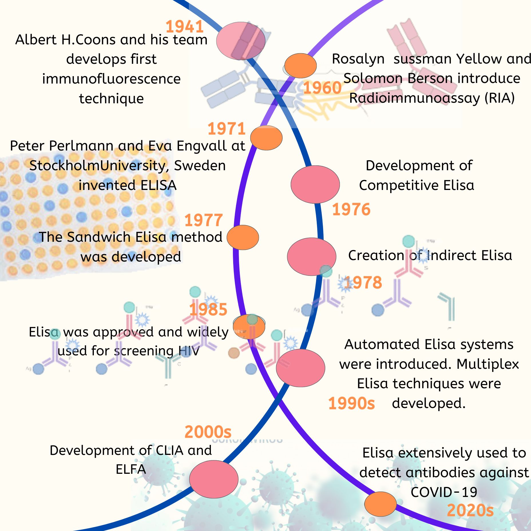
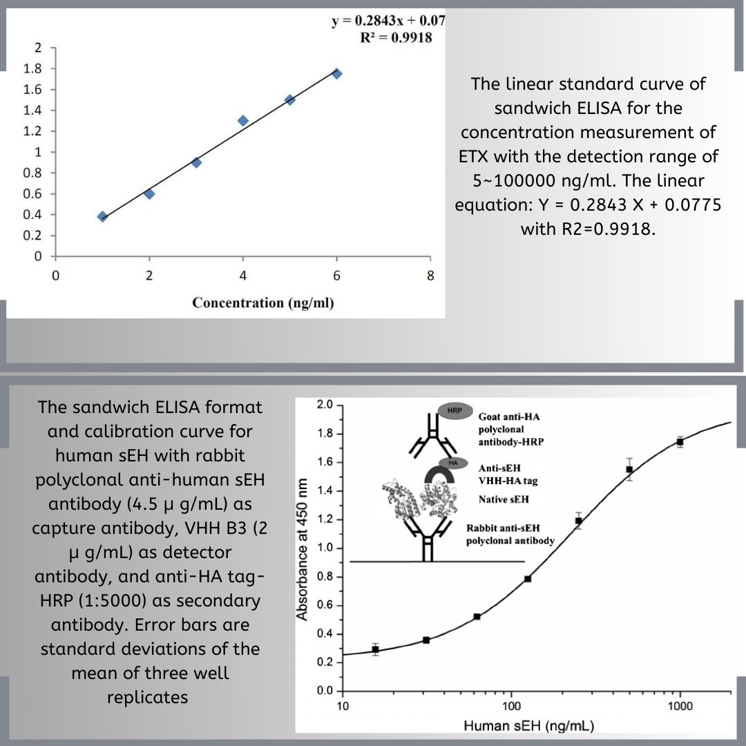
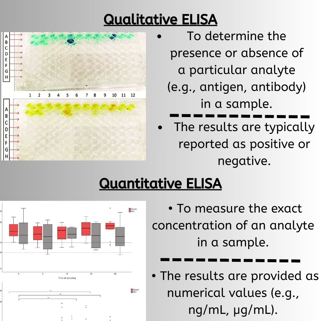
In ELISA, documentation is critical for tracking validation steps and ensuring consistency throughout the process. Proper labeling of samples and reagents is essential to avoid mix-ups. Using a calibrated pipette for accurate measurements helps in minimizing variability and pipetting errors, which are common in ELISA assays. Optimization of reagent concentrations and adherence to the protocol improves assay performance and reproducibility. For example, maintaining proper incubation times and room temperature conditions is necessary to achieve reliable results.
Proper sample preparation is very important because it affects how accurate and reliable the ELISA test is. This process includes steps to reduce interference, keep the sample intact, and create the best conditions for the test. For safety, different levels of protection are needed based on how contagious the materials are. Basic protective clothing helps prevent sample contamination and incorrect results. Essential safety gear includes a lab coat, gloves, eye protection, and sometimes a fume hood.

|
Storage of Samples: |
|
|
Sample Collection: |
|
|
Sample Preparation: |
|
|
Using Frozen Samples: |
|
Resource Optimization:
To enhance efficiency, it’s crucial to manage resources like assay plates, wash buffers, and reagents effectively. Employing a reliable plate washer and adhering to protocols for washing steps can significantly reduce background signals and improve sensitivity. Regular data analysis ensures that the assay performance meets expected standards, especially when dealing with low analyte concentrations.

|
Cell Culture Supernatant: |
|
|
Cell Extract: |
|
|
Conditioned Medium: |
|
|
Milk, Urine, Saliva: |
|
|
Plasma: |
|
|
Serum: |
|
|
Tissue Extract: |
|
|
General Recommendations: |
|
- Reagent Preparation and Quality Control: Proper reagent preparation and validation are key to achieving reliable assay performance. Consistent labeling and control of reagents, along with tracking usage and storage, contribute to the overall reliability of the ELISA procedure. Implementing stringent quality controls ensures that analyte concentrations are measured accurately and that results are reproducible.t 10,000 rpm for 5 minutes at 4°C before use.
|
Storage Duration Limits: |
|
|
Avoiding Repeated Freeze-Thaw Cycles: |
|
|
Storage of Kits: |
|
|
First In, First Out: |
|
|
Pre-Warming: |
|
|
Storage of Unused Components: |
|
Sample Processing
- In Elisa sample processing, centrifugation is used to remove cellular components and debris, which is important for getting clean supernatant.
- Further cleaning the samples by removing leftover particles through depth or membrane filtration methods. This step ensures the samples are free from any contaminants that could affect the test results.
Explore Our Full Range of ELISA Kits
Whether you’re testing human, animal, or plant samples, MyBioSource offers over 1 million ELISA kits covering thousands of analytes across every major species.
References
- Bloomer, R. J. (2015). Considerations in the Measurement of Testosterone in Saliva and Serum using ELISA Procedures. British Journal of Medicine and Medical Research, 5(1), 116-122.
- Besnard, L., Fabre, V., Fettig, M., Gousseinov, E., Kawakami, Y., Laroudie, N., … & Pattnaik, P. (2016). Clarification of vaccines: An overview of filter-based technology trends and best practices. Biotechnology advances, 34(1), 1-13.
- Parandakh, A., Ymbern, O., Jogia, W., Renault, J., Ng, A., & Juncker, D. (2023). 3D-printed capillaries ELISA-on-a-chip with aliquoting. Lab on a Chip, 23(6), 1547-1560.
- Sanjay Kumar, Yogesh Kumar, Dharam Malhotra, Shruti Dhar, Anil Nichani. Standardization and comparison of serial dilution and single dilution enzyme-linked immunosorbent assay (ELISA) using different antigenic preparations of the Babesia (Theileria) equi parasite. Veterinary Research, 2003, 34 (1), pp.71-83.
- Colella, A. P., Prakash, A., & Miklavcic, J. J. (2024). Homogenization and thermal processing reduce the concentration of extracellular vesicles in bovine milk. Food Science & Nutrition, 12(1), 131-140.
- Rey, G., Schuetz, F., Schroeder, D., Kaluschke, C., Wendeler, M. W., Hofmann, I., … & Obrdlik, P. (2024). Automated ELISA for potency measurements of therapeutic antibodies and antibody fragments. Journal of Pharmaceutical and Biomedical Analysis, 245, 116141.

