|
ELISA |
RIA |
|
|
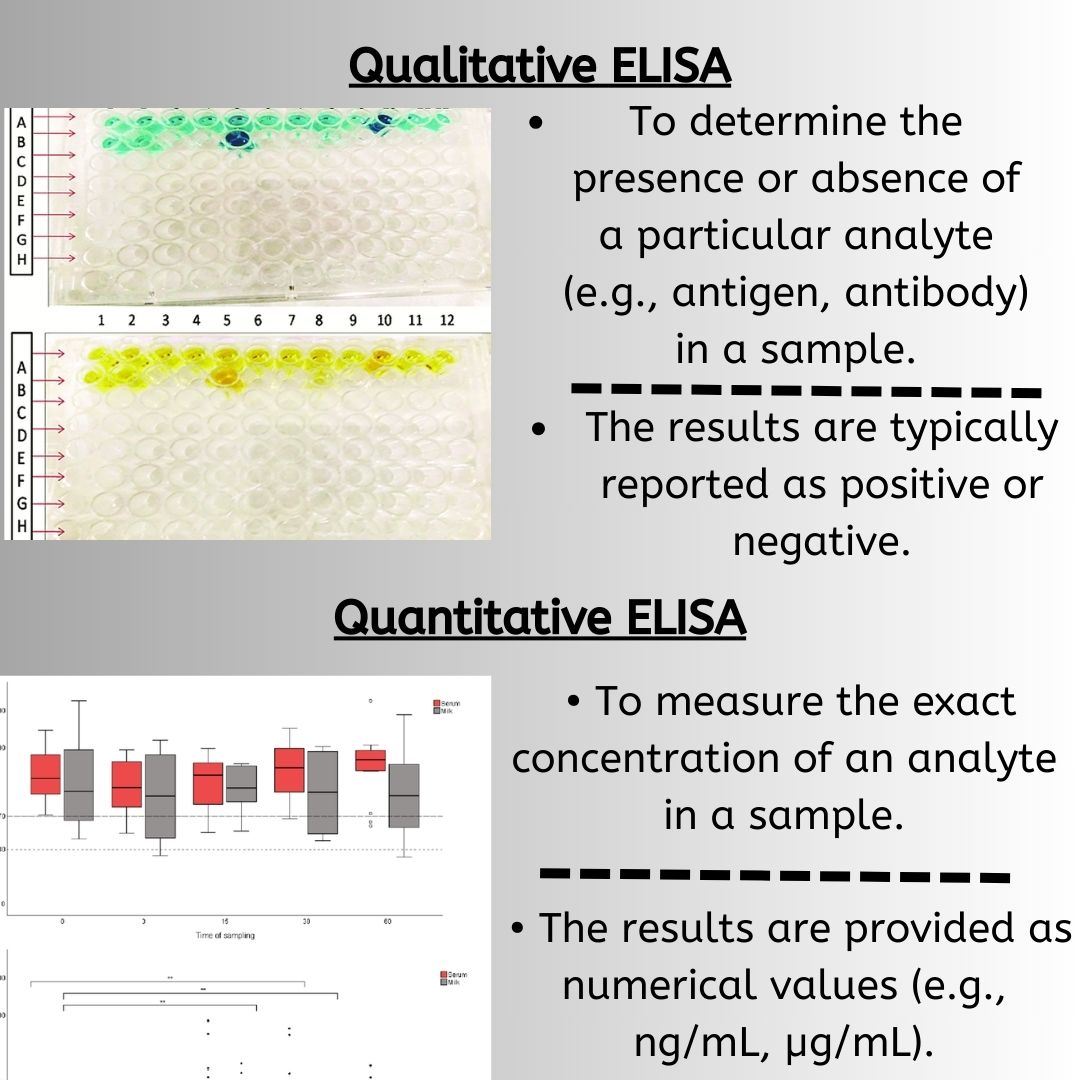
Basic Principle |
Uses enzymes linked to antibodies. Detection is based on a color change or light emission from an enzyme-substrate reaction. |
Uses radioisotopes to label antigens.
Detection is based on measuring radioactivity emitted from the antigen-antibody complex. |
|
Detection Mechanism Safety |
Produces a measurable color change or light emission. | Measures radioactivity emitted by the radioisotopes. |
|
Safety |
Safe, no radiation hazards.
No special handling or disposal requirements. |
Involves radiation hazards.
Requires careful handling, storage, and disposal of radioactive materials. Potential radiation hazards need to be documented and reported. |
|
Laboratory Requirements |
Can be conducted in standard laboratories.
Does not require specialized facilities or training. |
Requires specialized laboratory facilities for handling radioactive materials.
Needs dedicated areas and specialized training for personnel. |
|
Cost and Convenience |
Generally lower cost.
No specific arrangements for storage or disposal of materials. |
Higher cost due to the use of radioisotopes.
Requires meticulous planning for the handling, storage, and disposal of radioactive substances. |
|
Sensitivity |
High sensitivity, but generally less sensitive than RIA. | More sensitive than ELISA. |
|
Regulatory and Safety Concerns |
No specific regulatory issues beyond standard laboratory safety. | Strict regulatory and safety concerns due to the use of radioactive materials.
Short half-lives of isotopes limit its application. |
|
Fluoroimmuoassays |
Elisa |
|
|
Basic Principle |
Use fluorescent labels attached to antibodies.
Detection is based on the measurement of fluorescence intensity emitted by antigen-antibody complexes. |
Use enzyme-linked antibodies.
Detection is based on a color change or light emission from an enzyme-substrate reaction. |
|
Detection Mechanism |
Measure fluorescence intensity. | Measures color change or light emission. |
|
Sensitivity |
Highly sensitive, suitable for detecting low concentrations of antigens. | High sensitivity, but generally less sensitive than fluoroimmunoassays for very low concentrations. |
|
Applications |
Suitable for applications requiring high sensitivity.
Often used in research where low antigen concentrations are measured. |
Widely used in clinical diagnostics and research.
Versatile for detecting and quantifying antigens in various biological fluids and tissues. |
|
Ease of Use |
Requires equipment capable of detecting fluorescence. | Simpler to set up with standard lab equipment like a microplate reader. |
|
CLIA |
ELISA |
||
|
Basic Principle |
Uses molecules that emit light when returning from an excited state to a ground state.
Enzymes like alkaline phosphatase or horseradish peroxidase catalyze reactions with substrates (e.g., luminol), producing light. |
Uses enzyme-linked antibodies.
Detection is based on a color change or light emission from an enzyme-substrate reaction. |
|
|
Detection Mechanism |
Measures light emission. | Measures color change or light emission. | |
|
Sensitivity and Specificity |
Higher sensitivity and specificity compared to ELISA.
Shows higher sensitivity in detecting antibodies for diseases like SARS-CoV-2 (92.3% to 97.8% vs. ELISA’s 75.0% to 84.3%). |
High sensitivity and specificity, but generally less than CLIA. | |
|
Applications |
Used for detecting and quantifying antigens and antibodies for various diseases such as SARS-CoV-2, HIV, HCV, HBV, Mycoplasma pneumoniae, Hepatitis C virus, Treponema pallidum, and more. |
||
|
Ease of Use |
Provides rapid results.
Requires specialized equipment to measure light emission. |
Simpler to set up with standard lab equipment like a microplate reader.
More time-consuming compared to CLIA. |
|
|
Cost-Effectiveness |
Generally more expensive due to specialized equipment and reagents. | More cost-effective, making it suitable for routine use. | |
|
Detection Range |
Measures relative light units, offering a broader detection range. | Measures optical density, which can be more limited in range. | |
|
ELISA |
Western Blotting |
|
|
Basic Principle and Application |
Measures antigen-antibody reactions using optical density.
Preferred for its simplicity, speed, and suitability for high-throughput labs. Uses 96-well plates and requires lower sample volumes. Quantifies antigen concentration using a standard curve. |
More complex and time-consuming.
Requires careful sample loading, consideration of membrane and signal saturation, and use of internal controls for normalization. Offers high specificity and can detect protein size and purity. Capable of multiplex detection with fluorescently labeled antibodies. |
|
Ease of Use |
Simple to perform and suitable for large-scale testing. | Complex and requires more time and expertise.
Involves multiple steps including electrophoresis, transfer to membranes, and antibody detection. |
|
Sensitivity and Specificity |
High sensitivity and good specificity for detecting and quantifying antigens. | High specificity, with the ability to confirm the presence and purity of specific proteins.
Can detect the size of proteins, providing additional information about the target. |
|
Applications |
Widely used in clinical diagnostics and research.
Ideal for high-throughput screening and routine assays. |
Invaluable in research and clinical settings where detailed protein analysis is required.
Used for confirming specific targets and analyzing protein size and purity. |
|
Detection and Quantification |
Relies on optical density measurements.
Uses a standard curve for accurate concentration determination. |
Allows visualization of protein bands, providing information on protein size.
Can use internal controls for normalization to ensure accurate results. |
Explore Our Full Range of ELISA Kits
Whether you’re testing human, animal, or plant samples, MyBioSource offers over 1 million ELISA kits covering thousands of analytes across every major species.
References
- Muddasir Khan1M.Phil., et. Al, Enzyme-Linked Immunosorbent Assay versus Chemiluminescent Immunoassay: A General Overview
- Luong, K., Lozier, B. K., Novis, C. L., Smith, T. L., Zuromski, L. M., & Peterson, L. K. (2024). Comparison of three methods for the detection of antibodies against muscle-specific kinase. Journal of Immunological Methods, 526, 113627.
- Sletten, G. B., Løvberg, K. E., Moen, L. H., Skarpeid, H. J., & Egaas, E. (2005). A comparison of time-resolved fluoroimmunoassay and ELISA in the detection of casein in foodstuffs. Food and agricultural immunology, 16(3), 235-243.
- Owen, C., Fader, K. A., & Hassanein, M. (2024). Western Blotting: Evolution of an Old Analytical Method To a New Quantitative Tool for Biomarker Measurements. Bioanalysis, 16(5), 319–328.
- Saeed Ahmed, Jianan Ning, Dapeng Peng, Ting Chen, Ijaz Ahmad, Aashaq Ali, Zhixin Lei, Muhammad Abu bakr Shabbir, Guyue Cheng & Zonghui Yuan (2020) Current advances in immunoassays for the detection of antibiotics residues: a review, Food and Agricultural Immunology, 31:1, 268-290,