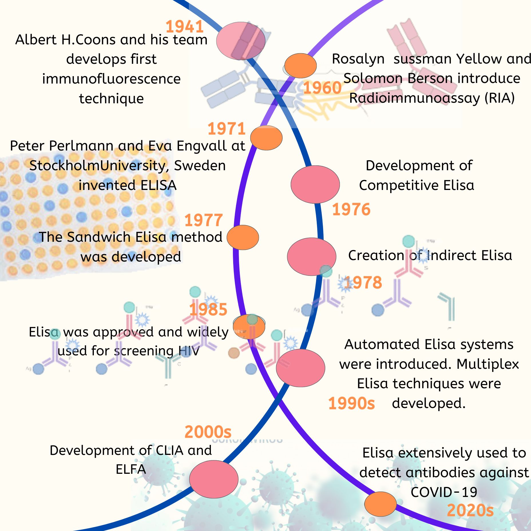
ELISA is a biochemical technique that utilizes antibodies and an enzyme-triggered color change to detect either antigens, such as proteins, hormones, and peptides, or antibodies in a provided sample. The “indirect” and “sandwich” methods of ELISA are capable of detecting antigens or antibodies at low concentrations. ELISAs come in various configurations, depending on their intended use. ELISA can be categorized into two primary types as a solid-phase method: competitive assays that employ either an antigen–enzyme conjugate or an antibody-enzyme conjugate, and non-competitive assays that use a double antibody “sandwich” technique where the second antibody has an indicator enzyme linked to it.
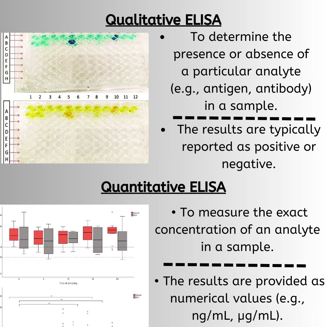
The most important step in the ELISA assay is to directly or indirectly identify the antigen by attaching or immobilizing either the antigen or the antigen-specific capture antibody onto the surface of the well. To achieve sensitive and reliable measurements, a “capture” antibody can selectively extract the antigen from a mixture of antigens in the sample. This results in the antigen being “sandwiched” between the capture antibody and the detection antibody. In situations where the antigen to be measured is small or has only one epitope for antibody binding, a competitive approach is employed. In this approach, either the antigen is labeled and competes for the formation of the unlabeled antigen-antibody complex or the antibody is labeled and competes for the antigen bound to the sample. Each of these modified ELISA techniques can be used for both qualitative and quantitative purposes.
The ELISA protocol involves several steps, which are outlined below:
-
Coating:
Coating is a critical step in which an antigen or capture antibody is immobilized onto a microtiter plate to create a solid-phase matrix. The antigen or antibody is typically coated onto the wells of the microtiter plate by incubating the plate with a solution containing the antigen or antibody. The choice of antigen or antibody to be coated onto the plate depends on the specific application and the target molecule to be detected. Proper optimization of the coating step, including the concentration and incubation time of the antigen or antibody, is essential to ensure the success of the assay.

-
Blocking:
Blocking is a step where non-specific binding sites on the microtiter plate are blocked to prevent non-specific binding of antibodies or other proteins to the plate. Blocking is essential to reduce background noise and improve the specificity of the assay. Blocking is typically done by adding a protein such as bovine serum albumin (BSA), casein, or milk to the wells after the plate has been coated with the primary antibody or antigen. The blocking protein binds to any remaining unoccupied sites on the plate, reducing the likelihood of non-specific binding of antibodies or other proteins to the plate.
The plate is then incubated to allow the blocking protein to bind to the plate, and any excess blocking protein is removed by washing the plate with a buffer solution. After blocking, the primary antibody or antigen can be added to the wells.
-
Sample and standard addition:
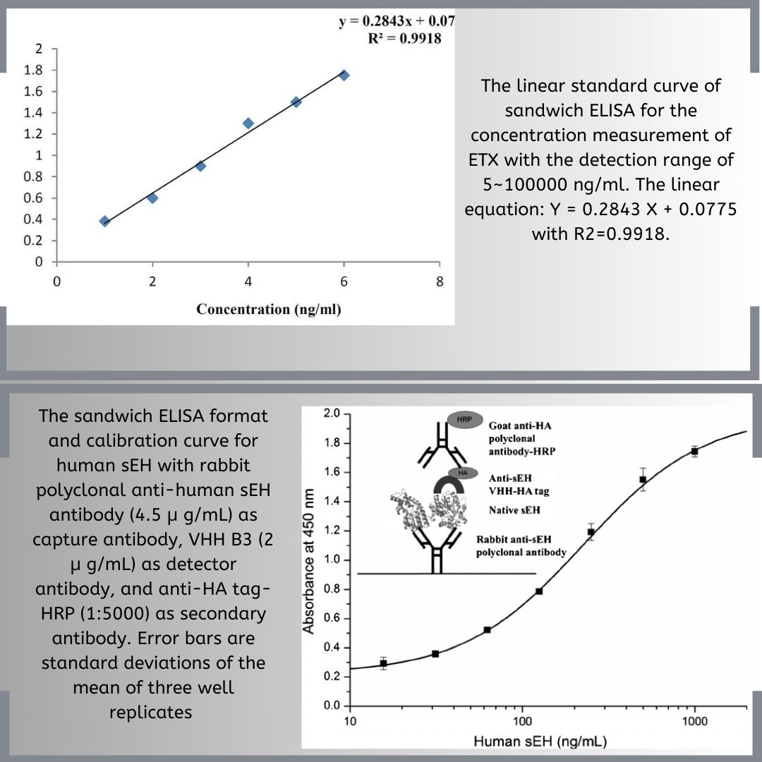
The standards are typically a set of known concentrations of the antigen that are used to generate a calibration curve, which is used to quantify the amount of the antigen in the unknown samples. The standards are added in known concentrations in separate wells of the plate, whereas the unknown samples are added in duplicate or triplicate wells.
-
Primary antibody incubation:
The primary antibody incubation step involves the addition of a specific antibody that recognizes and binds to the antigen of interest that has been coated onto the microtiter plate. The primary antibody is typically raised in a specific species, such as mouse or rabbit, and may be labeled or unlabeled. If the primary antibody is unlabeled, a secondary antibody conjugated to an enzyme such as horseradish peroxidase (HRP) or alkaline phosphatase (AP) is added in a later step to generate a measurable signal.

-
Secondary antibody incubation:
Secondary antibody incubation is a crucial step in the ELISA assay, where a secondary antibody is added to the microtiter plate to bind specifically to the primary antibody that was previously bound to the antigen or antibody coated on the plate. The primary antibody-antigen complex acts as a bridge to link the secondary antibody to the microtiter plate, allowing the enzyme to bind to the substrate and generate a measurable signal. The signal generated by the enzyme–conjugated secondary antibody is proportional to the amount of the primary antibody or antigen present in the sample.
-
Substrate addition:
In ELISA, a substrate is added to the microtiter plate after the incubation of the secondary antibody to generate a measurable signal. The substrate reacts with the enzyme–conjugated to the secondary antibody and produces a detectable signal that can be quantified using a microplate reader. The choice of substrate depends on the type of enzyme used, and the most commonly used substrates are chromogenic or chemiluminescent. Chromogenic substrates produce a colored reaction product that can be read at a specific wavelength, whereas chemiluminescent substrates produce light, which can be measured by a luminometer. The reaction time and substrate concentration should be optimized to generate an adequate signal without causing background noise.
-
Stop solution addition:
A stop solution is added to the microtiter plate to stop the enzymatic reaction between the substrate and the enzyme–conjugated secondary antibody. The stop solution changes the pH of the reaction mixture, which causes the enzymatic reaction to stop and prevents further color development or luminescence. A commonly used stop solution is sulfuric acid, which is added to the wells after the substrate incubation step. The addition of the stop solution is essential to ensure that the reaction does not continue and that the signal generated is stable and reproducible. After adding the stop solution, the absorbance or luminescence of each well is measured using a microplate reader.
-
Signal detection:
The signal is generated when the substrate is added to the microtiter plate and reacts with the enzyme to produce a colorimetric or chemiluminescent signal, depending on the substrate used. The signal generated is proportional to the amount of the primary antibody or antigen present in the sample. The results are analyzed using software or other statistical methods to determine the concentration of the primary antibody or antigen in the sample.
Conclusion
Each stage of the ELISA will influence the final result and therefore great care must be taken to optimize and then standardize the method. The most important influencing factors are antigen coating, choice of plates, choice of blocking agent, and choice of secondary antibodies and detection system. Each of these factors will vary with each antigen being used.
Overall, the ELISA protocol is highly sensitive and has numerous applications in research, clinical diagnosis, and drug development.
References
- (PDF) Enzyme Immunoassay and Enzyme-Linked Immunosorbent Assay (researchgate.net)
- Reen, D. J. (n.d.). Enzyme-Linked Immunosorbent Assay (ELISA). Basic Protein and Peptide Protocols, 461–466. doi:10.1385/0-89603-268-x:461
- https://link.springer.com/protocol/10.1385/1-59259-321-6:243
- Conceptual view of the ELISA assay. Streptavidin-coated (blue ovals)… | Download Scientific Diagram (researchgate.net)


